
for all enquiries please call today on
01924 635006
Email Enquiries
37 Years Personal Experience • Registered to practice by Environmental Health Department • Advanced Cosmetic Practitioner
Skin Blemish Identification
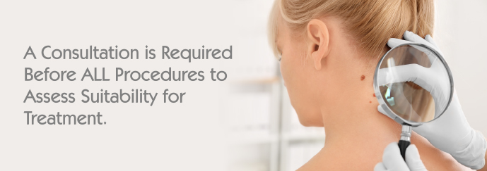
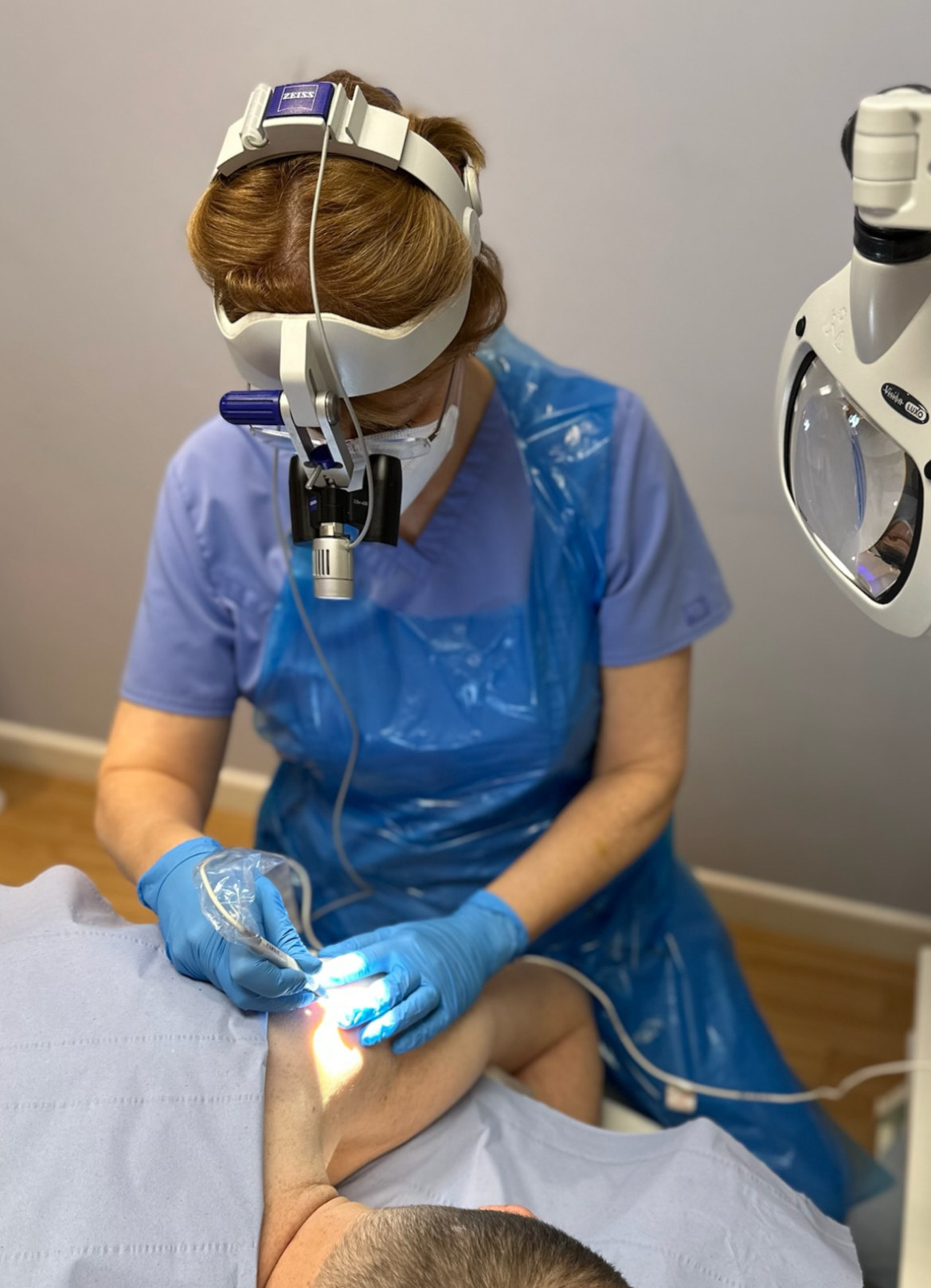
The following is a list of skin blemishes that are suitable for treatment with medically approved ‘Diathermy’ techniques. They can either be removed, reduced or improved, which will depend on the blemish and suitability for treatment. Assessment and correct identification are paramount for an effective outcome to be achieved. If you have a blemish of concern, then just make contact and I will be happy to check it for you. There are some examples of treatment results on the 'Gallery and Testimonial' Page.
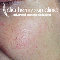 |
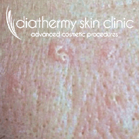
Red Veins (Telangiectasia) Commonly occur on nose, cheeks, chin and chest. Highly visible superficial dilated capillaries also known as ‘broken capillaries’, ‘thread veins’ or ‘broken veins.’ There are many causes, however, ageing, genetics, hormonal changes, medications, lifestyle choices (including smoking) and certain skin conditions are contributary factors. |
 |
Spider Neavi (Telangiectasia) Seen frequently on the nose, face and chest. Distinguished by a central dilated blood vessel with smaller vessels radiating from the centre point. Hence, they look like the legs of a spider from which it derives its name. The main cause is through some type of trauma to the skin but also some hormonal changes and medical conditions can also be responsible. |
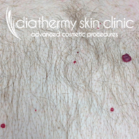 |
Blood Spots (Cherry Angiomas, Campbell de Morgan’s - CDM) Commonly seen on the torso, upper arms and legs and around the hair line of the face. They are a compact area of concentrated blood with a translucent skin layer, so appearing bright in colour. They may appear as a single blemish or in profusion. There exact reason for appearance is not really known. However, commonly appearing with age (35+), hormonal, medical or familial history. |
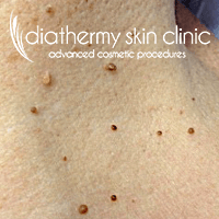 |
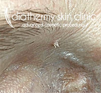
Skin Tags and Papillomas (Fibro Epithelial Polyps-Multiple) These are harmless fibrous skin growths, having a loose and ’wiggly’ appearance (pedunculated). Their size can be very small or increasing to be very large. Commonly found in areas of skin friction e.g., neck, underarms, midriff and groin. Causes can be attributed to skin folds and skin movement, weight gain and recent studies show a correlation to some subtypes of HPV – However, they are not contagious from person to person. |
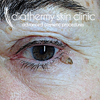 |
Skin Tag – Eye Eye polyps (tags) are commonly seen around the eyes, particularly as age progresses. A combination of movement of skin folds and loose skin can develop these blemishes. Diathermy is a very precise and effective way to safely remove them. |
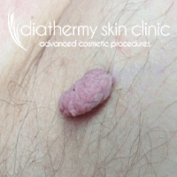 |
Acrochordon (Large Skin Tag or polyp) Singular large tags can sometimes be identified as ‘acrochordons’, which simply means that they are defined by a very thin stalk attachment to the skin and a large soft fibrous body. Commonly seen again in areas of friction, particularly under the arm, groin or behind the knee. |
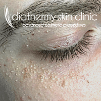 |
Milia Cysts Commonly seen around the cheekbones, eyes and cheeks (although not exclusively), they appear as white/cream firm, raised, pearl-like lumps. The contents are made up of compressed keratin from an undeveloped sebaceous or sweat glands with a blind duct. Some common causes thought to be responsible are dry skin conditions (although not exclusively), familial history, incorrect use and choice of skin products including excessive use of vitamin C and environmental conditions. |
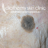 |
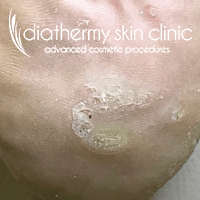
Seborrhoeic Keratosis (Seborrhoeic Wart) These blemishes vary in size and colour from skin coloured through to black. Having defined edges, cracked, rough or waxy surface they give a ‘stuck on’ appearance! Some can also contain milia cysts! Commonly seen on the torso, face and limbs. Although sometimes called a ‘skin wart’, they are actually not warts at all and not contagious. The most recognised cause is the ageing process particularly on skins with a familial predisposition. |
 |
Dermatosis Papulosa Nigra (DPN) These blemishes are ‘histologically’ the same as seborrhoeic keratosis but do not scale or crust. They are smooth, domed and hyperpigmented. They occur commonly on darker skin types and ethnicities and are seen around the cheeks, temples, forehead and neck. They respond very well to the gentle precise techniques used with Diathermy. |
 |
Sebaceous Hyperplasia Commonly seen on the forehead, cheeks and chest it is a condition affecting the sebaceous glands. They can be singular or multiple and appear as creamy/yellow plaques of trapped sebum and often seen with a central pore area or punctum. The blemishes often develop creating ‘lobules’ which give the appearance of a ‘doughnut or cauliflower’ effect. They can often look worse through slight swelling after physical activity. |
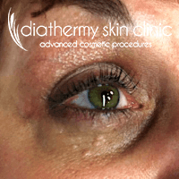 |
Syringoma These blemishes affect mostly the lower eye tissue although can be seen on the upper lid too. There is also a version of this blemish that appears on other parts of the body too. They are harmless sweat gland tumours which appear slightly raised, smooth and generally creamy in colour. The cause is not exactly known but is thought that hereditary factors, oilier skin or in people living in hotter, humid climates. |
 |
Xanthelasma Palpebrarum (Xanthomata) These soft, yellowy plaques are found in symmetry on the upper and lower eyelids and less commonly on other parts of the body. They are associated with lipid metabolism and can sometimes be referred to as cholesterol deposits. It is recommended that to establish causation medical assessment should be sought, as there are several underlying reasons for their appearance. The plaques themselves can be irritating but not harmful. |
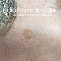 |
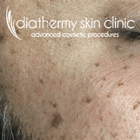
Age Spots (Liver Spot/Chloasma Disc/Solar Lentigo) Often seen on the face, chest and hands. These are harmless, macular discolorations and tend to appear with age. However, repeated sun exposure is also a common cause. Their defined colour is formed from the build-up of ‘lipofuscin’, which accumulates in the connective tissue of the skin. Hence, they have a nickname of ‘sunspots or brown spots’ |
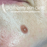 |
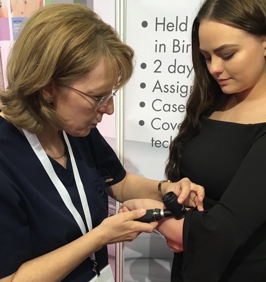
Mole (Pigmented and Non-Pigmented) REDUCED ONLY! The number of moles on a person’s face and/or body can vary greatly from one to several hundred! They may appear pigmented or non-pigmented. Raised moles are generally smooth and may also contain strong deep hairs, which can be treated separately with electrolysis. It is very important that the history and characteristics of ‘any’ mole are identified before treatment is deemed suitable. Medical referral may be advised. Dermoscopy will be used during this process to determine the moles' suitability for treatment. Raised moles are ‘only’ dehydrated to reduce their size and not actually removed! |
 |
Common Wart & Plane Warts Common Warts are seen as raised firm growths with rough crusty appearance, often described as a ‘cauliflower’ appearance. The majority being caused by the HPV virus. Although many may self-resolve, some can remain for years and become painful or appear unsightly. They are contagious with direct viral contact. Plane Warts – Small flesh coloured or pigmented blemishes, they may look slightly raised with a dry surface. Commonly seen on the forehead, cheeks and chest and also associated with subtypes of the HPV virus. |
 |
Filiform Wart Quite distinctive in appearance as they are raised from the skin with narrow ‘frond like’ protrusions. Commonly affecting the neck, eye, chin,nose and lip areas. There attachment is soft and responds very well to Diathermy treatment. |
 |
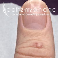
Verruca Pedis (Singular and Mosaic) Also known as ‘plantar wart’. Appearing on the feet as compressed warts. Quite often black spots can be seen in the blemish which are thrombosed capillaries. They can be very painful to walk on and although can self-resolve they can be in situ for many years. Caused by the HPV virus and are contagious with direct viral contact. Careful hygiene is needed to avoid their spread both locally on the foot and to others. |
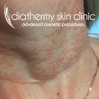 |
Poikiloderma of Civatte A compounded area of skin discolouration affecting the neck and décolleté area commonly forming a ‘T-Shirt’ pattern. Developing mainly from over exposure to sunlight, although genetics can also have an impact. Mottled reddening of the area is its main feature although the skin can be occasional raised and a little rough to touch. A combination of treatment techniques is used to reduce its appearance together with continued sun protection. |
| Other treatable conditions | Epidermoid Cysts – Raised, sac contains ‘cheesy’ odorous contents. Sebaceous Cysts – Raised, oily filled sac, non-odorous. Pilar Cysts – Firm moveable lumps on scalp enclosed strong sac. Fibrous and Granular Blemishes. Raised firm non-pigmented lumps. |


ACP Practitioner: Mrs Janet Turner Lecturer, Assessor, Cert Ed. Queens Drive Ossett.
Telephone: 01924 635006
Site designed and built by PAB Studios Ltd
Telephone: 01924 635006
Site designed and built by PAB Studios Ltd